
Sleep study reports come in a lot of variety. Some reports will be “just the facts.” Others will have TONS of data. Here are the things that are important for us to review as dentists, and what we should point out to our patients.
So why do we care? I believe it is important to show the patient what is going on with them so that they better understand their problem.
Is it an in lab test, a home sleep test, or are you looking at the CPAP titration?
The first thing you need to determine is what type of study report you are looking at. You want to be looking at a baseline study, not the CPAP (continuous positive airway pressure) titration. Here are the different types of studies:
- Inlab polysomnogram, or PSG. This is the gold standard test and is performed with the patient going to a sleep lab, getting wired up, and spending the night in the lab while a technician attends the study.
- A “home sleep test” or “out of center sleep test” (OCST). These are small devices that are usually sent home with the patient or sometimes mailed to them. They usually consist of a pulse-oximeter, 1 or 2 strain gauges, and a nasal cannula. Some home test units can also measure brainwaves or have another means to determine if the patient is actually asleep or not (most home units only assume the patient is asleep…making them less accurate).
- A “split night study.” This is an inlab PSG where the first part of the nightis the diagnostic phase, and then, IF the patient shows significant sleep apnea, the patient is awoken and placed on CPAP. The rest of the night is used to find the optimal CPAP pressure to treat the sleep apnea.
- CPAP titration study. This is an inlab PSG where the entire night is used to adjust the CPAP. Usually in these reports there will be an initial sentence about what the baseline PSG showed.
Once you know that you are looking at the baseline PSG or OCST, here are the things you want to look for:
- AHI: What was the overall Apnea/Hypopnea Index (AHI)? The AHI is the measure of how bad the patient’s sleep apnea is. The scale of AHI is:
- < 5 = normal in an adult. (In a child > 1.5 indicates clinically significant sleep apnea)
- 5-15 = mild
- 15-30 = moderate
- > 30 = severe
Now, let’s take this AHI number and break it down a bit. Obstructive apneas and central apneas are added together to get the “A” in the AHI. Central sleep apnea is where the brain doesn’t tell the person to try to breathe. [As a brief note, pure central sleep apnea is very, very rare, BUT if you ever see a patient with a high percentage of central sleep apnea, instead of obstructive, you will want to review the goals of oral appliance therapy with their physician, as oral appliance therapy typically will not affect central sleep apnea — neither does CPAP.] Hypopneas are the “H.” A hypopnea is a reduction in ventilation by at least 50% that also results in a decrease of the O2 saturation by 4% or more. In other words, a hypopnea is shallow breathing that results in desaturation. These are usually obstructive in nature, like partly kinking a hose, but not completely blocking the flow.
I enjoy showing the patient the difference between an apnea and a hypopnea by drawing on the back of one of the forms. I say, “in the sleep lab, or in your home study, you had a nasal cannula in your nose. Normally when you see these things they are to give someone extra oxygen. In this case, the cannula was measuring your breathing in and out. On the computer screen it would look like this:
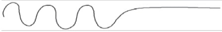
I’ll tell the patient that this, an apnea, happened “X number of times” throughout the whole night. This number will usually, but not always, be in the report. This is not the “index” but the actual number of obstructive and central apneas that occurred throughout the night. If the number of apneas is not specified in the report, then you can’t show this.
I will then show the patient what a hypopnea would look like on the computer screen in the sleep lab:
 I’ll tell the patient that this, a hypopnea, occurred “X number of times” throughout the whole night.
By showing the patient the difference between an apnea and a hypopnea, it helps them to understand their problem better, and makes the severity of their sleep apnea make more sense… because the AHI does not tell the whole story.
Speaking of story, here’s a little mathematical story problem for you:
Patient A’s study shows that he had 60 obstructive apneas throughout the night. He had 30 hypopneas throughout the night. He slept 6 hours total. Therefore his AHI is (60 + 30) / 6 = 15.
Patient B’s study shows that he had 30 obstructive apneas throughout the night. He had 60 hypopneas throughout the night. He slept 6 hours total. Therefore his AHI is (30 + 60) / 6 = 15.
Wait a second! They both have an AHI of 15 even though one had half as many actual episodes of stopping breathing? Yep!
Now let’s take this to the extreme. What is the AHI if the patient had 180 apneas for the night, 0 hypopneas for the night, and slept 6 hours? (180 + 0) / 6 = 30.
What is the AHI if the patient had 0 apneas for the night (literally NEVER stopped breathing), 180 hypopneas for the night, and slept 6 hours? (0 + 180) / 6 = 30.
What? So BOTH of these patients have “severe sleep apnea,” even though the second one NEVER stopped breathing?! That’s correct.
So why do we care? I believe it is important to show the patient what is going on with them so that they better understand their problem. If you are told that you have severe sleep apnea and that you stop breathing 30 times per hour, but your wife of 20 years says that she has only rarely noticed you stopped breathing, are you going to believe the report? Probably not. So it is important to explain to the patient that even though they have been told that they “stop breathing X times per hour” (which is what they will think the AHI is) that they don’t actually completely stop breathing all of those times (unless of course they have 0 hypopneas throughout the night).
For some of our patients you will be the first one to go over the baseline sleep study with them.
It’s also important for us to look at this as I believe, through experience, that we tend to have an easier time treating patients with more hypopneas than apneas. That doesn’t mean that we don’t treat people with lots of apneas, but it just means that we might “lower their expectations” a little of oral appliance therapy completely resolving their apneas.
- Sleep Position: In conjunction with the AHI you will also usually find information about sleep position and the AHI when the patient is sleeping supine versus on their side. For most people their obstructive sleep apnea is worse on their back (supine). For some people you will notice that their problem almost exclusively occurs when they sleep supine. When you notice this, you should talk to the patient about this fact and encourage them to sleep as much as possible on their side, including once they get their oral appliance.
- O2 Saturation: What is the O2 saturation nadir (lowest point), and how much time did the patient spend with an O2 saturation below 90%?
This is a pretty obvious one to us as to why it is important. However, most patients will not realize what the O2 saturation means. Explain to them that our blood O2 levels, at this elevation, should be above 95% most of the time. Explain that if they were in a hospital and their O2 level went below 90, alarms would go off! Then tell them that their O2 level dropped to a low of X and was below 90 X% of the night.
- Sleep Stages: How much time the patient spent in the different levels of sleep during the study. Non REM sleep stages are referred to as N1, N2 and N3. Here are the “ideal” percentages:
- N1 is “light sleep” or “transitional sleep.” This should only account for about 5-10% of the total sleep time.
- N2 is “restful sleep.” This should be about 45-55% of the total sleep time. When people have reduced deep sleep and REM sleep, they usually have increased N1 and N2 sleep.
- N3 is “deep sleep” or “slow wave sleep.” This should be about 10-20% (much more in children, and becomes less as we get older).
REM is Rapid Eye Movement sleep, or “dreamsleep.” We should have about 20-25% of our sleep be REM sleep. In REM sleep the muscles have much less tone (some will say paralyzed), and as such obstructive sleep apnea tends to be worse in REM sleep.
While there are a lot of things that are fascinating about how sleep works, here are the simple things you need to know and share with your patients.
First, if they have reduced deep sleep (N3) they will feel physically tired. They may also have muscle pain, or even “fibromyalgia” type symptoms.
Second, if they have reduced REM sleep they will feel mentally tired. They may also have memory problems and a “clouded intellect.”
For some of our patients you will be the first one to go over the baseline sleep study with them. For many of our patients it was months or years ago that their doctor reviewed their sleep study with them, so they have likely forgotten much of the information. Going over this information with the patient will help them, and you, to understand their problem much better and make them, in my opinion, more likely to stick with treatment.
Each sleep lab and sleep doctor will present their data a little different, but you should be able to find the above information in all sleep studies and help the patient to understand it.
We DO NOT base our appliance selection on any of this information. ALL oral appliances work the same way… they keep the mandible from falling back, or keep it slightly forward. The data WILL help us to know how bad the patient’s obstructive sleep apnea is so that we will better know how to treat them and how important it will be for them to return to their physician for objective follow up and adjustment of the oral appliance in the sleep lab.
Follow Up Sleep Study
I believe that ALL patients should be referred back to the referring physician (the one who wrote the prescription for the oral appliance) for consideration of a follow up sleep study with the oral appliance in place. IF the physician does decide to have a follow up sleep study, I also believe that it is ideal to have the appliance adjusted in the sleep lab by the sleep techs (you will normally need to teach them how to do this and have written protocols for this).
When comparing a baseline study to a follow up study, make sure that you compare apples to apples, and look for:
- PSG to a PSG is apples to apples, but
- How long has it been since the last PSG?
- Were both studies at the same lab?
- Were both studies read by the same doctor?
- PSG to HST, or HST to PSG = not apples to apples = tough to make conclusions
- HST to HST maybe apples to apples, but
- How long since the last HST?
- Is the same HST device being used (if not, probably not apples to apples)?
Once you understand the differences between the technical aspects of the baseline study versus the follow up study, look for the following things that may be different from the baseline study to the follow up study:
- How long has it been since the baseline study? Sleep apnea usually gets worse as we get older.
- Has there been any weight gain? Sleep apnea usually gets worse with weight gain.
- Different sleep posture? Sleep apnea is usually worse in the supine position.
- Look at more than just the AHI
- Was there a change in the number of apneas?
- Was there a change in the number of hypopneas?
- Was there a change in the average O2 saturation? The nadir?
I have had several patients that prior to me referring them back to the physician for consideration of a follow up sleep study with adjustment of the appliance in the sleep lab, the patient reported feeling fantastic and having a major improvement of their snoring. However, when they went in for the follow up sleep study the report came back that they didn’t do as well as I would have liked. In almost all of these cases I was able to compare the baseline study to the follow up study and find the reasons that we didn’t see a big change in the AHI, even though the patient felt much better. The most common things I’ve seen that made the follow up study numbers not as good as I would have liked were:
- It had been 5 or more years since the baseline study.
- The patient had gained significant weight.
- The patient slept mostly non-supine on their baseline study, and mostly on their back on their follow up study.
The bottom-line is that it is important for us as dentists to understand what is presented in sleep study reports AND when follow up studies are completed to make sure that we compare the follow up study to the baseline study, and make sure that our objective data appears to be consistent with the subjective data of what the patient is reporting to us.