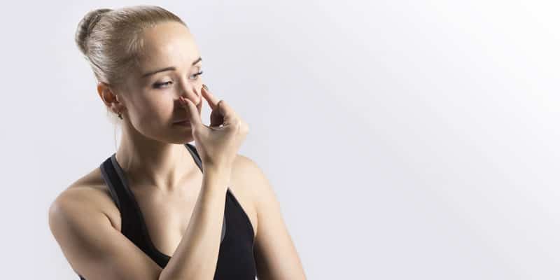
Patrick McKeown explains how breathing re-education can be beneficial, taking into consideration the connections between breathing volume, CO2 tolerance, oral breathing, and nervous system balance.
 by Patrick McKeown
In the last decade, the field of sleep medicine has changed radically. Sleep apnea was previously considered a purely anatomical issue, but we now know this is not the case. Research has identified three non-anatomical phenotypes, signifying a combination of contributing factors. Upper airway collapsibility and craniofacial anatomy are, of course, still relevant, but the underlying cause of sleep apnea differs from one patient to another.
A problem exists in the treatment of sleep apnea. Traditional CPAP treatment, which focuses on the anatomy, is subject to poor compliance and often fails to fully resolve the condition. Therapies such as weight loss, electrical stimulation of the tongue, and treatment of nasal obstruction with steroids, surgery, or airway stents, also focus on anatomical correction. However, an understanding of the traits that contribute to sleep apnea indicates an underlying issue at the root of symptoms. One that none of these methods address: Dysfunctional breathing.
The Root Cause of Sleep Apnea
The four phenotypes of sleep apnea, defined by Eckert et al. in 2013, are pharyngeal critical closing pressure (Pcrit), loop gain, upper airway recruitment, and arousal threshold. Later research further refines this concept into one of four endotypes, facilitating a model of personalized treatment.
Pcrit
Pcrit is used to measure the collapsibility of the airway in sleep-disordered breathing. It represents the pressure of negative suction required to close the airway during sleep. Contributing factors include airway narrowing and collapsibility due to dysfunction in the airway dilator muscles. A narrow airway increases resistance, making it more vulnerable to collapse. Fat around the pharynx, torso, and abdomen, all restrict function of the upper airway. This is exacerbated when breathing is hard and fast, especially through an open mouth. The apnea/hypopnea is characterized by a drop in airflow, but these events are often heralded by excess breathing volume. After each apnea, the patient resumes breathing with a large gasp, perpetuating further apneic events.
Pcrit is closely related to oral breathing. When the mouth is open, the upper airway is more vulnerable to collapse, independent of any nasal obstruction, and even of breathing route. This is due to mechanical obstruction caused by upper airway narrowing, and inefficient contraction of upper airway dilator muscles. Mouth breathing is linked with greater oxygen desaturation and can also cause CPAP non-compliance.
During nasal breathing, the tongue can sit in its correct resting place in the upper palate and is less likely to block the airway. Nasal breathing is also important for adequate diaphragm excursion, which supports lung volume, and helps keep the airway open. Regular practice of diaphragm breathing exercises can improve Pcrit by enhancing the strength of the respiratory tract and improving the organization of breathing from the central nervous system.
Often, after nasal surgery or adenotonsillectomy in children, symptoms return because the habit of persistent oral breathing is not addressed. Therefore, restoration of nasal breathing, day, and night, must be the first step in breathing re-education. My own clinical experience indicates that the only way to ensure nasal breathing during sleep is to use supports such as paper tape across the lips, chin up strips or MyoTape®.
Loop Gain
Loop gain reflects chemosensitivity to carbon dioxide (CO2). Patients with high loop gain have an excessive physiological response to small changes in CO2. This is directly related to low breath-hold time. Indeed, chemosensitivity can be easily determined using breath-hold time after exhalation – a measurement fundamental to breathing re-education.
During an apnea, breathing stops. CO2 is unable to leave the body via the lungs and builds up in the bloodstream. Because CO2 provides the primary stimulus to breathe, an increase in the pressure of CO2 of just 2-5mmHg can more than double ventilation. When breathing re-starts after an apnea, a patient with high loop gain will experience exaggerated breathing in response to small changes in CO2. This can trigger hyperventilation, inhibiting the respiratory signals and causing a central apnea to occur. At the same time, unstable breathing contributes to airway collapse, producing an obstructive apnea. There is evidence that some sleep apnea patients with high loop gain during sleep are also highly sensitive to changes in CO2 while awake.
Around 30% of sleep apnea patients have high loop gain. As this is a non-anatomical trait, mandibular advancement devices do not tend to be effective. However, the chemosensitivity to CO2 and low breath-hold time synonymous with loop gain can be improved using breathing exercises that normalize breathing rate and minute ventilation.
Upper Airway Recruitment
This refers to the mechanical efficiency of the upper airway dilator muscles. The human pharynx lacks rigid, bony support, but there are more than 20 muscles in the upper airway. These muscles are involved in respiratory and non-respiratory functions, and their activation counters the negative suction pressure created during inhalation. Depending on the dynamic balance between negative suction pressure and neural drive to the upper airway dilator muscles, the airway can be vulnerable to collapse during sleep.
It is time to get to the root cause of symptoms in sleep apnea treatment.
Upper airway recruitment threshold is defined by the level of stimulus required to activate the upper airway dilator muscles. When the muscles do not respond well to airway muscle collapse, the severity of sleep apnea can increase.
Patients with sleep apnea tend to have poor control of the upper airway dilator muscles during inhalation, whether they are awake or asleep. They also tend to have weaker airway muscles.
Breathing re-education includes exercises that improve the strength and function of the breathing muscles, especially the diaphragm. It also restores the proper resting posture of the tongue. Tongue position is important in sleep apnea, as the genioglossus muscle in the tongue plays a role in maintaining an open airway. For oral breathers, it can be helpful to re-educate the tongue muscles and improve tone and function in the upper airways using Myofunctional Therapy (MT). MT can restore nasal breathing during sleep – a marker of successful upper airway treatment. It also reduces snoring and improves CPAP compliance and adherence. Moreover, nasal breathing harnesses the gas nitric oxide, which helps maintain tone in the airway dilator muscles.
Arousal Threshold
Arousal threshold reflects whether the patient is a light or deep sleeper. This is defined by the levels of airway pressure and change in arterial CO2 concentration required to wake the patient. Those with a low arousal threshold and poor upper airway recruitment will awaken before the dilator muscles have activated. They will experience frequent, unnecessary arousals, sleep fragmentation and daytime fatigue. Low arousal threshold is linked to insomnia, autonomic imbalance, and mood disorders. What’s more, men and women with low arousal threshold are at the greatest risk of all-cause mortality.
When the upper airway muscles do not work properly, sleep that is too deep can also be problematic. If the patient fails to awaken during an apnea, breathing can stop for longer, causing significant and damaging oxygen desaturation.
Nasal breathing supports deeper sleep, lowering arousal threshold. It slows the breathing rate. Conversely, heightened ventilation induces arousal from sleep, regardless of its cause. This is one reason low arousal threshold and insomnia often coexist. Slow, nasal breathing activates the parasympathetic nervous system via the vagus nerve, while mouth breathing, which is often fast and into the upper chest, is associated with the stress response. Those patients with chronic stress and high anxiety struggle to fall and stay asleep. Breathing exercises that involve a respiratory rate of six breaths per minute optimize parasympathetic tone and reduce stress, ensuring deeper, more restful sleep with fewer arousals.
The Four Phenotypes and Breathing Re-Education
Given the connections between breathing volume, CO2 tolerance, oral breathing, and nervous system balance, it makes sense to address the root cause of symptoms on an individual basis, using the breath.
Breathing re-education restores functional breathing from three dimensions: biochemical, biomechanical, and resonant frequency (cadence), with a foundation of full-time nasal breathing. Nasal breathing is vital for dental health, but it can also support treatment of all four phenotypes of sleep apnea, increasing the chance of fully resolving the condition.
There is, as yet, limited research into the relationship between dysfunctional breathing and sleep apnea. However, successful novel approaches involving breathing control have included myofunctional therapy, wind instrument and didgeridoo playing, diaphragm breathing, singing exercises and the Buteyko Breathing Method.

Key Takeaway
Breathing re-education has substantial potential benefits for patients with sleep apnea. The goal should always be to reach a comfortable breath-hold time after exhalation of 25 seconds. Nocturnal mouth taping is effective, but it is not enough. Daytime breathing must be functional too.
It is time to get to the root cause of symptoms in sleep apnea treatment and find a way to fully resolve the condition. A simple program of breathing exercises offers an accessible, personalized approach.
You can find out more about your patients and tailor their rebreathing education by asking the right questions. Read “Patient Education!” by Glennine Varga to find out more about how to connect with patients. https://dentalsleeppractice.com/patient-education/
 Patrick McKeown is a leading authority in the field of breathing for health, performance, and sleep. He was educated at Trinity College Dublin, completing his clinical training in Russia. In 2002, he was accredited as a breathing coach by renowned physician, Dr. Konstantin Buteyko. Patrick is a Fellow of the Royal Society of Biology, creator and Master Instructor at Oxygen Advantage®, and founder of Buteyko Clinic International, the leading Buteyko breathing clinic in the UK. For the last 20 years, he has taught functional breathing to help children and adults with asthma, sleep apnea, and many health conditions. Patrick’s 2021 book, “The Breathing Cure,” contains comprehensive research into breathing for sleep, and offers practical solutions for chronic conditions. His article, “Breathing Re-Education and Phenotypes of Sleep Apnea: A Review,” co-authored with Drs. Carlos O’Connor-Reina and Guillermo Plaza, is published in the Journal of Clinical Medicine.