by Cara Riek, DNP, RN, FNP-BC, IBCLC, DABLS; Stevanie Bahnerth, DC; and Peter Vitruk, PhD, MInstP, CPhys
The goal of frenectomy is to release the restricted frena (i.e., Tethered Oral Tissues or TOTs). The goal of a Functional Frenectomy1-3 in children, adolescents and adults is to release the restricted frena and to restore, with the help of mandatory pre- and post-frenectomy Oro-Myofunctional Therapy (OMT), the mobility and functionality of tongue and lips for optimal breathing, speech, chewing, swallow, and posture.1-3 A Functional Frenectomy involves, besides the surgeon, a team trained in myofunctional, physical, craniosacral, osteopathic, and chiropractic therapies.
This article reports clinical cases of a Functional Frenectomy with the CO2 laser and includes, in addition to pre- and post-frenectomy OMT, a pre- and post-surgical systematic bodywork performed by a chiropractor, to achieve long-lasting functional results.
CO2 Laser Functional Frenectomy approach incorporates the following three parts:
- Pre-surgical chiropractor assessment, bodywork and OMT exercises to strengthen and re-pattern tongue function;
- CO2 laser frenectomy with real-time assessment of tongue and lip restrictions by the clinician to achieve ideal release for the optimal function;
- Post-frenectomy chiropractor assessment, bodywork and OMT exercises to ensure long-lasting functional results.
The compensations developed by the muscles can create muscle tension and tightness that need to be addressed before frenectomy, i.e., patient may require pre-frenectomy care to help begin to loosen and align the joints affected by the TOTs. Such collaboration between frenectomy provider and chiropractor may successfully resolve chronic pain, headaches, airway obstruction, and digestive problems.
Case Studies
The Functional Frenectomy approach to releasing oral restrictions is illustrated by the clinical cases in Figures 1-2.
Patient 1

The 9 year-old female patient, presented with an open bite (Figure 1A), a narrow, high palate (Figure 1B) and limited range of mouth opening (Figure 1C). She wanted to eliminate the habit of persistent thumb sucking that she found embarrassing. The patient was started on MyoBrace appliance and was undergoing chiropractic care. The patient was unable to keep the MyoBrace in most of the night. The patient’s mother attempted mouth taping, but the patient often pulled the tape off in her sleep. While the patient was improving, she hit a plateau and the progress slowed down. During that time the patient was diagnosed, by Dr. Riek, to have a posterior tongue-tie.

After undergoing OMT, the patient’s lingual and labial (not pictured) restriction was released by Dr. Riek with a LightScalpel CO2 laser. A combination of topical and local anesthetic was used. Figure 1D shows immediate post-op image of the tongue – note lack of bleeding and clean surgical margins; no sutures were needed.

Post-frenectomy, OMT continued and a much improved tongue mobility was achieved as tongue healed (Figures 1E and 1F: both at six months post revision. The tongue range of motion ratio (TRMR) has increased from 32/46 mm to 48/50 mm.
Post-frenectomy, the patient now easily retains the MyoBrace appliance in the mouth without the need to tape. She no longer sucks on her thumb. Post-frenectomy, a significant progress was observed in both the palate shape and the bite – see sequences in Figures 1A and 1B.
Patient 2
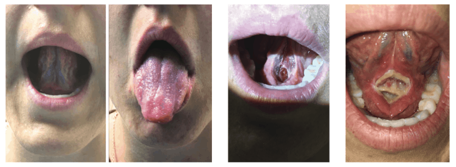
Figure 2A Figure 2B Figure 2C
The 28 y. o. female patient came to Dr. Riek’s office with complaints of headaches, neck pain, not getting restful night sleep, and digestive concerns. Upon examination, a tongue-tie was noted (figure 2A shows the limited range of tongue motion). Dr. Riek recommended OMT four weeks prior to the functional frenum release and the continuation of chiropractic care.
After the pre-frenectomy care, the patient returned to Dr. Riek’s office for her tongue-tie release procedure. The local anesthesia was administered. The same LightScalpel CO2 laser settings were utilized as for Patient 1. Figure 2B shows the patient immediately after the laser release. No bleeding occurred intraoperatively or postoperatively. The wound, albeit deep, was left to heal by secondary intention. The surgical site healed without complications (figure 2C shows the wound healing five days following laser frenectomy). In the absence of notable pain and swelling, the patient was able to perform her OMT exercises without difficulties.

Figure 2D Figure 2E
Nine months post laser frenectomy and ongoing OMT and chiropractic care, the patient has a much improved tongue range of motion: at the initial visit to Dr. Riek, the patient’s TRMR was Grade 2 (36/50); post-procedure the TRMR increased to 52/55) (Figure 2D). In addition, the lift of the tongue has significantly improved (Figure 2E). The initial symptoms have been resolved. The patient started mouth taping at night to achieve better sleep and reports no night time waking. Her headaches are minimal now. Neck pain was completely resolved, she is able to sleep 8 hours per night WITHOUT waking, and no longer experiences GERD (swallowing air, some reflux, burping).
Why CO2 laser?
Not all lasers are equally good at vaporizing (i.e., ablating or cutting) and coagulating soft tissue. Figure 3 demonstrates the difference in the absorption spectra for the main soft tissue chromophores for different laser wavelengths.4 The CO2 lasers offer the following benefits for oral soft tissue surgery:
- Approximately 1,000 times greater photo-thermal cutting efficiency relative to dental diodes, and in approximately 10 times greater photo-thermal coagulation/hemostasis depth relative to erbium lasers;
- Close match between the coagulation depth of the CO2 laser and the blood capillary diameters (Figure 3).4 This close match distinguishes CO2 from erbium lasers and provides instant hemostasis during high speed ablation or cutting. The CO2 laser ablates tissue while coagulating small blood and lymphatic vasculature. It provides improved visibility of the surgical field and therefore enables more precise and accurate tissue removal;
- Minimal post-operative edema, pain and discomfort; due to the intraoperative closure of lymphatic vessels on the margins of the CO2 laser incision.4 With CO2 laser frenectomy, patients report less post-operative pain and discomfort than with the scalpel.

CO2 Laser Frenectomy Settings
For both clinical cases described above, the LightScalpel CO2 laser was set to 2 W Non-SuperPulse, and gated at 20 Hz, 40-60% duty cycle, 0.8-1.2 W average power; straight tipless handpiece with a 0.25 mm focal spot diameter was used to produce 0.3-0.4 mm deep shallow incisions at a 3-5 mm/sec hand speed. Such shallow incision depth, combined with shallow coagulation depth (0.1-0.2 mm4), allows for excellent and progressive visualization of larger diameter blood vessels. Also due to the shallow depth of incision, multiple laser passes are required to complete the required depth of incision in a safe and controlled fashion.
Summary
Achieving, with the help of OMT, the optimal breathing, speech, chewing, swallowing and posture is limited in the presence of restricted frena (i.e., Tethered Oral Tissues or TOTs).1-3 Such limitations are removed by a Functional Frenectomy, which includes mandatory pre- and post-frenectomy OMT. Pre- and post-frenectomy evaluation and bodywork by a chiropractor are beneficial additions to Functional Frenectomy as they help achieving truly long-lasting functional results. The use of CO2 laser for frenectomy results in a less painful surgical site, than the conventional procedure. The clinician can use either topical or small amounts of local anesthesia for functional tongue and lip assessments during the procedure.
Acknowledgments: Authors greatly appreciate the help and contribution from Anna (Anya) Glazkova, PhD, in preparing this material for publication.
Read more about how clinicians use the CO2 laser in functional frenectomy in “Functional Frenectomy (Osteopathically Guided)” here.